Thursday, 27 December 2018

Thyroid hormonesT3 and T4|their formation and secretion

Thyroid hormones-formation and secretion


Thyroid mainly secretes two major hormones T3 and T4, also called as throxine and triiodothyronine respectively.Both of these hormones increase metabolic rate of the body. Thyroid secretion of these two hormones is mainly controlled by thyroid stimulating hormone(TSH), which is secreted by anterior pituitary gland.

Formation of thyroid hormones

About 50 milligrams of ingested iodine in the form of iodide is required for the formation of thyroxine. Iodides from the diet are absorbed into the blood from GIT tract and from their are selectively remove by the cells of thyroid gland for the formation of thyroid hormones.
The following steps explain the synthesis of thyroid hormones:

Iodide pump

  •  The transport of iodides from blood into thyroid cells or follicles is accomplished by the basal membrane of thyroid cells, which has the ability to transport iodides to interior of cell,through active transport.
  • The sodium-iodide symporter, which co-transport one iodide along with two sodium ions through plasma membrane to the interior of cell 
  • Sodium potassium ATPase pump provides energy for the transport of iodide against concentration gradient, and also establish the low intracellular sodium concentration and gradient for facilitated diffusion of sodium into the cell by pumping sodium out of the cell.
  • This overall process of concentrating the iodide in the cell is called iodide trapping, which is influenced by several factors one of which is.the concentration of TSH.
  • Chloride iodide ion counter transport molecule called pendrin, helps in the transportation of iodide through the thyroid cell across the apical membrane into the thyroid follicles
Oxidation of iodide ion

  • After iodide trapping into the thyroid cells,the thyroid ions are converted to an oxidized form of iodine either nascent or I3,the purpose of this step is to make the iodide ion capable of combining directly with the amino acid tyrosine.
  • Amino acid tyrosine (Each molecule of thyroglobulin, a glycoprotein molecule secreted by thyroid epithelial cells into follicles,contain about 70  tyrosine amino acids and they are the major substrates that combine with the iodine to form thyroid hormones)
  • Enzyme peroxidase promote the oxidation of iodine,which is attached to the apical membrane of  the cell,thus providing the oxidized iodine at exactly the point where thyroglobulin molecule,after synthesis comes out from the golgi apparartus and through the cell membrane into stored thyroid cell colloid.

Organification of thyroglobulin

  • The binding of iodine with the thyroglobulin is called the organification of thyroglobulin.
  • The oxidised iodine will bind directly to the tyrosine of thyroglobulin to form thyroid hormones,first tyrosine is iodized to monoiodotyrosine, and then to triiodotyrosine.
  • Then in few days more and more iodotyrosine coupled with one another
  • when two molecules of diiodothyronine are joined together, the major hormonal product thyroxine is formed
 

Secretion of thyroid hormones from thyroid gland

  • First, the thyroxine and triiodothronine is cleaved from thyroglobulin and then release from the gland
  • This cleavage is accomplished when the apical surface of thyroid cells sends out pseudupod extensions that close around small portion of colloids to form pinolytic vesicles.
  • Then lysosomes in the cell cytoplasm fuse with these vesicles to form digestive vesicles which contain digestive enzymes from lysosomes mixed with colloid.
  • Multiple proteases digest the thyroglobulin and release thyroxine and teriiodothyronine in free form 
  • In this way thyroid hormones released into the blood.
The hypothalamus produces TSH releasing hormone that signals the pituitary to release TSH which stimulate thyroid to release thyroid hormones.

Wednesday, 26 December 2018

Overview anatomy of thyroid gland|location,Relations and blood supply

Thyroid Gland Anatomy

Thyroid is a large ductless gland that secretes hormones that control metabolism.It is located in the base of your neck below the Adam's apple,situated between C5 and T1 vertebrae.It is a butterfly shaped structure that is divided into two lobes, that are connected by isthmus.


Relations of thyroid gland

It has two borders and three surfaces
  • Anterior and posterior borders
  • Medial,lateral and post-lateral surfaces
Lateral surface 
It is covered by 
  • Sternohyoid
  • superior belly of omohyoid
  • sternothyroid
  • Anterior border of sternocleidomastoid 
Medial surface
  • Trachea and oesophagus
Postlateral surface
  • carotid sheath and overlaps common carotid artery
Anterior border

  • Anterior branch of superior thyroid artery 
Posterior border

  • Parathyroid glands
  • On left side thoracic duct

Muscles

The muscles associated with thyroid gland are
  • Sternocleidomastoid muscle
  • Sternothyroid muscle
  • Sternohyoid muscle
  • Sternocricoid muscle

Membranes

It has two membranes associated with it
  • Thyrohyoid membrane
  • Cricothyroid membrane

anterior neck muscle

    BLOOD SUPPLY


    Artery
    • Carotid Artery
    • Superior and inferior thyroid arteries
    Veins
    • Superior,middle and inferior thyroid veins
    • Jugular vein

    NERVE SUPPLY

    • Vagus nerve
    • cervical ganglion

    Physiological anatomy of thyroid gland

    • Thyroid gland is composed of a large number of closed follicles(100 to 300 micrometer in diameter)filled with large number of secretory substance called colloid.
    • And is lined with cuboid epithelial cells which secretes into the follicle, a glycoprotein called thyroglobulin,which contains the thyroid hormones.
    anatomy and physiology


    Tuesday, 25 December 2018

    Nephron physiology|How urine is produced actually by nephron

    Nephron Physiology-Urine formation

    In renal system filtration the blood is filtered by nephrons to produce urine, which is waste byproduct contains metabolic wastes and water.The rate at which different substances excreted in the urine is the sum of three renal processes;
    1. Glomerular filtration
    2. Reabsorption of substances from the renal tubules into the blood
    3. Secretions of substances from the blood into renal tubules
    Urine formation begins when a large amount of fluid that is virtually free of protein is filtered from the glomerular capillaries into the bowman's capsule
    Let's discuss the three renal processes by which urine is actually formed.

    Gromerular Filtration

    • The mechanism by which glomerulus filters the blood, supplied by afferent arteriole to glomerulus,is based upon high glomerular hydrostatic pressure,which forces blood to leave the glomerulus through efferent arteriole resulting in the filtration of blood.
    • Like most capillaries, the glomerular capillaries are relatively impermeable to proteins,so the filtered fluid is usually free of protein
    • There are few low-molecular wight substances, such as calcium and fatty acids , that are not freely filtered because they are partially bound to plasma proteins
    • The filtrate now contains large amount of water,glucose, aminoacids,urea, uric acid, electrolytes,the concentrations of which are similar to the concentrations in the plasma.
    • The volume of filtrate formed by both kidneys each minute is called glomerular filtration rate, which is about 125ml/min when the net filtration pressure is 10mmHg.

    Tubular Reabsorption

    In reabsorption useful substances like most of water and solutes are reabsorbed to blood.
    • Selective reabsorption occurs in renal tubules.
    • As the filtrate moves to PCT,most of the water,organic nutrients such as glucose,amino acids, vitamins etc, are selectively reabsorbed into the blood.
    • After reabsorption of  substances through PCT,in which some substances reabsorbed passively and some through active transport,only 60-70 % filtrate reaches loop of henle.
    • Mostly sodium and chloride and some quantity of water absorbed in the loop of henle and the remaining filtrate moves to the DCT.
    • Mostly sodium is reabsorbed here, with some electrolytes,so the filtrate entering the collecting duct is quite dilute
    • The collecting ducts absorbs much water as the body needs

    Tubular Secretion

    • Tubular secretion plays an important role in determining the amount of potassium and hydrogen ions and a few other substances that are excreted in the urine
    • Certain foreign substances  and drugs are poorly reabsorbed  but in addition are secreted from the blood into the tubules ,so their excretion rate is high.
    • Excess k+ ion is secreted in the tubules and in exchange Na+ ion is reabsorbed to maintain homoeostasis
    • Hydrogen ion secretion is very important in maintaining normal blood pH.
    Now the fluid is mainly consist of water, urea, uric acid and inorganic salts which are constituents of urine.The diagram below demonstrate all steps mentioned above;

    wikimedia commons


    Monday, 24 December 2018

    All about nephron anatomy|The functional unit of kidney

    Nephron-the functional unit of kidney

    The microscopic structural and functional unit of kidney that actually produces urine in the process called excretion.Each kidney in the human contains about 800,000 to 1,000,000 nephrons.
    The kidney cannot regenerate new nephrons. Therefore, with renal injury, disease,or normal aging there is gradual decrease in nephron number.This loss is not life threatening because adaptive changes in the remaining nephrons allow them to excrete the proper amount of wastes,water and electrolytes.

    Nephron Anatomy

    Nephron is basically consists of two parts:
    • Renal corpuscle
    • Renal tubule
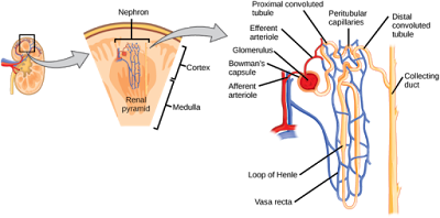
    Renal corpuscle consists of a tuft of capillaries called glomerulus encased in a cup shaped bowman's capsule.
    Renal tubules consists of proximal convulated tubule,loop of henle(ascending and descending loop of henle),distal convulated tubule and the collecting duct.
    • The glomerulus contains a network of branching capillaries that arises from afferent arterioles and empties into efferent arteriole.The glomerular capillaries have high hydrostatic pressure (about 60mmHg) due to smaller diameter of efferent arteriole than afferent arteriole.

    Glomerular Capillary Membrane

    The glomerular capillary membrane is similar to that of other capillaries,except that it has three instead of two major layers
    • The endothelium of the capillary with minute pores called fenestrations
    • A middle basement membrane
    • Layer of epithelial cells(podocytes)surrounding the outer surface of capillary basement membrane
    Together these layers make up the filtration barrier,which filters several hundred times  as much wastes as the usual capillary membrane.Even with this high rate of filtration, the glomerular capillary membrane normally prevents filtration of plasma proteins.
    CNX OpenStax
    • Bowman's capsule is a cup like structure which encloses the glomerulus.
    • Proximal convulated tubule-lies in the cortex of kidney and is the proximal part of the renal tubule,next to bowman's capsule.Maximum reabsorption of essential elements like water,glucose,aminoacids and electolytes takes place here.
    • From the proximal tubule fluid flows into  loop of henle,which dips in renal medulla.It is U-Shaped and consists of ascending and descending limb.The walls of the descending limb and the lower end of the ascending limb are very thin therefore are called the thin segment of loop of henle.After the ascending limb of the loop returns back to the cortex, its wall become much thicker,and thus called as thick segment of loop of henle.
    • Distal convulated tubule is similar in fuction with proximal tubule(reabsorption), and is the distal part of the renal tubule.
    • Collecting duct form by many collecting tubules. In each kidney there are about 250 very large collecting ducts,each collects urine from about 4000 nephrons and empties into minor then major calyx and then into renal pelvis.

    Types of nephrons

    Although each nephron has all the components described earlier, there are some differences,depending on how deep the nephron lies within the kidney mass
    • Cortical nephrons-they have glomeruli located in the outer cortex and have short loop of henle.For the cortical nephrons the entire tubular system is surrounded by  an extensive network of peritubular capillaries. 
    • Juxtamedullary nephron-they have their glomeruli lie deep in the cortex near the medulla,the vascular structures supply them also differs from those that supply cortical nephrons.

    Location, structure ,blood supply -A detailed overview of the anatomy of stomach

    Stomach:

    The stomach is the dilated portion of alimentary canal.Oesophagus connects the stomach with pharynx.

    Location:

    It is situated in the upper part of abdomen just below the oesophagus.

    Structure:

    It is roughly J shaped.It has two openings, two curvatures and two surfaces.

    • Openings: Cardiac and Pyloric orifice
    • Curvatures: Greater and Lesser curvature
    • Surfaces: anterior and Posterior surfaces

    The stomach is divided into:

    Fundus, body, pyloric antrum and pylorus.


    • Fundus is dome shaped and it is usually full of gas.
    • Body extends from the cardiac orifice to a constant notch in the lower part of lesser curvature(cisura angularis)
    • Pyloric antrum extends from cisura angularis to the pylorus
    • Pylorus has pyloric canal which is the cavity of the pylorus and pyloric sphincter.


    The lesser curvature forms the right border of stomach and is connected to liver and the greater curvature extends from the left of cardiac orifice  along the left border of stomach.

    The cardiac sphincter is present between oesophagus and stomach,it prevents the back flow of  stomach content into stomach.
    The pyloric orifice is forms by the pyloric canal. The circular muscles are much thicker here and forms the which prevents the back flow of intestinal contents to stomach.

    Blood supply:

    Arteries:

    • Right and left gastric arteries
    • Right and left gastropiploic arteries
    • short gastric arteries derived from the splenic artery.
    These arteries supply the lesser curvature, greater curvature and fundus respectively.

    Veins:

    • right and left gastric veins drains into portal vein.
    • short gastric and left gastropiploic vein drains into splenic vein
    • right gastropiploic vein drains into SMV(superior mesenteric vein).

    Nerve supply:

    • Sympathetic nerve supply is from the celiac plexus.
    • Parasympathetic is from the vagus nerve(10th cranial nerve).


    Saturday, 22 December 2018

    Urinary system|brief description|check anatomy and clinical notes about urinary system obstruction


    Urinary system

    Urinary system consist of a pair of kidneys,ureters and bladder and urethra.Its function is to eliminate wastes from the body.regulate blood volume,pressure and blood pH,it also helps synthesize calcitrol,regulates electrolytes and metabolites.
    Let's discuss one by one the elements or organs in urinary system.

    Kidney

    There is a pair of kidneys that lie behind the peritoneum high up on the posterior abdominal wall on either side of vertebral column. It is a bean shaped organ and has an outer cortex which is dark brown in colour and the inner medulla is light brown.The medulla is composed of approximately 12 renal pyramids,each having its base oriented towards the cortex and its apex(the renal papilla) projecting medially.The cortex projects into medulla between adjacent pyramids as renal columns.
    On the medial concave border of each kidney is the hilus,which extends into a large cavity (the renal sinus),The hilus transmits the renal pelvis, renal artery,renal vein and sympathetic nerve fibers.
    Major and Minor calyces
    Within the renal sinus,the upper expanded end of the ureter (renal pelvis)divides into two or three major calyces,each of which in turn divides into two  or three minor calyces
    The kidney has the four coverings which are:

    • Fibrous capsule-present on the outer surface
    • Perirenal fat-covers the fibrous capsule
    • Renal fascia-present outside the perirenal fat and also covers suprarenal glands along with kidneys.
    • Pararenal fat-external to renal fascia.


    Blood Supply

    Artery
    • Renal artery,branch of aorta 
    Vein
    • Renal vein drain into inferior vena cava.

    Ureters

    The two ureters are muscular tubes, each measures about 10 inches in length extends from the kidney to the posterior surface of bladder.Ureters carry urine from kidneys to bladder.Renal pelvis is its upper extended end.
    Clinical Findings
    Ureteric stones may be arrested at the following sites

    • where the renal pelvis joins the ureter
    • where the ureter is kinked as it crosses the pelvic brim to enter the pelvis
    • where the ureter pierces the bladder wall.

    Blood supply

    Arteries

    • Upper end-renal artery
    • Middle portion-The testicular or the ovarian artery
    • Inferior end-The superior vesical artery
    Veins

    • renal vein drains into inferior vena cava

    Bladder


    It is pyramidal in shape or tetrahedral, when empty and becomes ovoid when filled with urine.It has an apex,base, superior surface,two inferolateral surfaces and four borders(anterior,posterior and two lateral)
    It is present immediately behind the pelvic bones behind the pubic symphysis and in front of rectum in the female.When the bladder is empty it lies entirely within the lesser pelvis but when it becomes distended with urine, it expands upward and forward into the abdominal cavity.

    Relations


    • Apex connected to the umbilicus by median umbilical ligament
    • superolateral angle joined by ureters
    • inferior angle give rise to urethra
    • base or posterior surface is triangular,rectovesicle pouch in male and vesico uterine pouch in the female
    In adult male the capacity of bladder to store urine is from 120 to 320ml. The mean capacity is about 220ml.An amount of urine beyond 220ml causes a desire to micturate.
    Ligaments
    The ligaments of the bladder are of two types:
    True ligaments
    • lateral ligaments(right and left)
    • Puboprostatic ligaments(two on lateral and medial side)
    • posterior ligament(right and left)
    False ligaments
    • Anteriorly there are three folds
    • Laterally a pair of false lateral ligaments
    • Posteriorly a pair of false posterior ligaments

    Blood supply

    Arteries
    • Superior and inferior vesical arteries
    • Obturator and inferior gluteal arteries
    • Uterine and vaginal arteries in female
    Veins
    Vesical venous plexus passes backward in the posterior ligaments of the urinarybladder to drain into the internal illiac veins.
    Urethra
    The urethra is a narrow fibromuscular
     tube that connects the urinary bladder to the urinary meatus.Its function is the removal or transport of urine outside the body.In males, the urethra travels through the penis and in female the urethra connects to the urinary meatus above the vagina.

    Blood supply

    Arteries
    • Inferior vesical artery
    • Middle rectal artery
    • Internal pudendal artery
    Veins
    • Inferior vesical vein
    • Middle rectal vein
    • Internal pudendal vein

    Obstruction of the urethra

    Obstruction of the urethra causes urinary retention. Conditions such as prostatic hyperplasia, cystocele, .rectocele,constipation,urinary tract stones,urethral stricture can cause an obstruction. 

    Friday, 21 December 2018

    Pancreas Anatomy and Functions|check overview location,structure,ducts, blood and nerve supply

    Pancreas:

    The pancreas is both an exocrine and endocrine gland. It is the elongated structure.It is one of the accessory organ of the gastrointestinal system.

    Location:

    it is situated behind the stomach and peritoneum, lies on posterior abdominal wall.Its right side is in contact with small intestine and left side with the hilum of spleen.
    Parts of pancreas:
    Pancreas is divided into four parts:
    Head:
    It lies in the concavity of  C-shaped duodenum and it is disc shaped and is the widest part of pancreas
    Tail:
    The tail is the last part and extends to the hilum of spleen.
    Neck:
    Neck connects the head to the body,it is next to head,narrow and lies in front of the beginning of the portal vein
    Body
    The body passes upward and to the left across the midline, it connects neck to the tail.
      Uncinate process-the projection from the lower part of the head behind the superior mesenteric vessels.


    Exocrine function of Pancreas-the portion of the pancreas that makes and secretes digestive enzymes into the duodenum. This includes acinar and duct cells with associated connective tissue, vessels, and nerves. The exocrine components comprise more than 95% of the pancreatic mass.
    Endocrine function of pancreas- the portions of the pancreas (the islets) that make and secrete insulin, glucagon, somatostatin and pancreatic polypeptide into the blood. Islets comprise 1-2% of pancreatic mass. It regulates blood sugur.

    Duct system

    Main pancreatic duct along with bile duct opens into second part of major duodenal papilla of duodenum.
    The accessory duct drains the upper part of duodenum on the minor duodenal papilla.
    Blausen.com staff


    Blood supply

    Artrial supply
    • splenic artery
    • inferior pancreaticoduodenal arteries
    Venous supply
    Pancreatic veins drains into portal vein.
    Nerve supply
    Both sympathetic and parasympathetic fibers from celiac plexus supply the pancreas.

    Thursday, 20 December 2018

    Anatomy of liver,eight lobes of liver,grooves and fissures|blood and nerve supplies to liver

    Liver:

    The largest organ in the body and is the accessory organ of the gastrointestinal system. I t is completely covered by by a fibrous capsule but is only partially covered by peritoneum.

    Location: 

    The liver is predominantly located in the right hypochondrium and epigastric areas, and extends into the left hypochondrium.
    It occupies the upper part of abdominal cavity just beneath the diaphragm.It is usually 15 to 17 cm in length with its upper border usually at the level of nipples and lower border at the level of costal cartilage of the 8-9th rib.

    The  liver may be divided into a large right lobe and the small left lobe by the attachment of the peritoneum of the falciform ligament.
    The right lobe is further divided into:
    Quadrate lobe and caudate lobe.


    Couinaud's Anatomy:

    It is becoming the universal nomenclature for hepatic lesion localisation.It is based on portal segments.The liver has eight segments and each segment has its own blood supply and these are of both functional and pathological importance.
    Segment 1 is the caudate lobe,2 and 3 are the  left superior and inferior lateral segments respectively.Segment 4 is the medial segment of the left lobe and further divided into the 4(a) and 4(b). The right lobe consists of four segments.
    Segment 5 and 6 are the inferior anterior and posterior segments respectively.Segment 7 is the posterior superior segment and segment 8 is the superior anterior one.


    Fissures:

    The two fissures, fissure for the ligamentum teres and fissure for ligamentum vesnosum are present in the liver.

    Fissure for the ligamentum teres:

    The fissure that contains the ligamentum teres(the fibrous remains of the umbilicak vein) lies between the left lobe and the quadrate lobe.

    Fissure for ligamentum venosum:

    The fissure that contains the ligamentum venosum(fibrous remains of ductus venosus) lies between the left lobe and caudate lobe.

    Groove:

    The liver contains a groove for the inferior vena cava which lies between the right lobe  and caudate lobe. Here the hepatic vein joins the inferior vena cava.

    Ligaments of liver:

    There are various ligaments that attaches the liver to near structures, these are:
    • Falciform ligament :
    The falciform ligement is a two layered fold of peritoneum that attaches the liver to the diaphragm above and to the anterior abdominal wall below.It has a sickle shaped free margin that contains the ligamentum teres.
    • Coronary ligament (anterior and posterior folds):
      It attaches the superior surface of the liver to the inferior surface of the diaphragm and demarcates the bare area of the liver The anterior and posterior folds unite to form the triangular ligaments on the right and left lobes of the liver.
    • Triangular ligaments (left and right):
      • The left triangular ligament is formed by the union of the anterior and posterior layers of the coronary ligament at the apex of the liver and attaches the left lobe of the liver to the diaphragm.
      • The right triangular ligament is formed in a similar fashion adjacent to the bare area and attaches the right lobe of the liver to the diaphragm.
    • Lesser omentum – Attaches the liver to the lesser curvature of the stomach and first part of the duodenum. It consists of the hepatoduodenal ligament (extends from the duodenum to the liver) and the hepatogastric ligament (extends from the stomach to the liver). The hepatoduodenal ligament surrounds the portal triad.

    Blood supply of liver:

    Arterial supply

    • The celiac artery branches into hepatic artery.
    • Hepatic artery divides into left and right terminal branches that supplies the porta hepatis.

    Venous supply

    • Portal vein divides inro right and left terminal branches that enter the porta hepatis.
    • Three or more hepatic veins drain into inferior vena cava emerge from the posterior surface of liver.

    Lymphatic drainage:

    • celiac nodes
    • posterior mediastinal nodes 
    The above nodes receives the lymph drainage that passes from porta hepatis and diaphragm respectively to enter the nodes.

    Nerve supply

    The liver is supplied by both sympathetic and parasympathetic fibers from celiac plexus.
    The 10th cranial nerve(vagus nerve)gives off a large hepatic branch that travels to liver.